IUCN/SSC Otter Specialist Group Bulletin

©IUCN/SCC Otter Specialist Group
Volume 40 Issue 2 (April 2023)
Citation: Barbosa, P.M.L., Esteves, P.S., and Carvalho-Junior, O. (2023). (2023). Anatomical Study of the Conus Medullaris in the Neotropical River Otter (Lontra longicaudis). IUCN Otter Spec. Group Bull. 40 (2): 109 - 115
Anatomical Study of the Conus Medullaris in the Neotropical River Otter (Lontra longicaudis)
Procassia M.L. Barbosa1, 2, Pricila S. Esteves2, and Oldemar de Oliveira Carvalho-Junior2
1Universidade Federal de Santa Catarina; Departamento de Zootecnista e Desenvolvimento Rural, Rod. Admar Gonzaga, s/n, Itacorubi-SC
2Instituto Ekko Brasil, Servidão Euclides João Alves, S/N, Lagoa do Peri, 88066-000, Florianópolis, SC, Brazil
*Correspondance Email: atendimento@ekkobrasil.org.br
Received 18th May 2022, accepted 2nd March 2023
Abstract: The morphology and physiology of the Lontra longicaudis is not well understood, making it sometimes difficult to understand its anatomy or behavior. A descriptive anatomical study of the spinal cord of neotropical river otters is presented. It was conducted at the Wild Animal Research Laboratory at the Federal University of Santa Catarina, Brazil. Three female otters were used, two cubs and one adult, from the Instituto Ekko Brasil (IEB) — Otter Project. To analyze the spinal cord and its components, measurements of body length and epiaxial musculature and vertebral arches were registered from the cervicothoracic transition to the base of the tail. The conus medullaris reached the fourth lumbar vertebra. In one animal, it reached the fifth lumbar vertebra. At the level of the sixth lumbar vertebra, no spinal cord segments were observed. In neotropical river otters, the space between the sacrum and the first caudal (sacrococcygeal) vertebra can be used for epidural anesthesia injection. Practitioners should consider this description when considering injection sides for epidural anesthesia.
Keywords: physiology; morphology; carnivorous; Mustelidae
INTRODUCTION
The neotropical otter, Lontra longicaudis, one of the four species of otters present in South America, occupies a wide region that goes from Mexico to Uruguay. It is a carnivorous animal with aquatic habits, which belongs to the Mustelidae family (Wilson and Reeder, 2005). In addition, this otter species is known to being dangerously threatened in the Atlantic Forest biome (Rodrigues et al., 2013; Carvalho Junior et al., 2021). Little is known about the physiology of the species. It is present throughout the Brazilian national territory, but is rarely seen in nature due to twilight habits and solitary behavior (Carvalho Junior et al., 2010).
The morphology and physiology of the neotropical river otter are not well understood, making it sometimes difficult to understand its anatomy or behavior. A study on the vascularization of the aortic arch of the species, for example, observes that the origin of these vessels is similar to that described for domestic and wild carnivores (Barbosa et al., 2021). These results demonstrate that the aortic artery is the main vessel that distributes arterial blood to all systems, bringing oxygen and nutrients to different areas of the body, and collecting the carbon dioxide that results from cellular metabolites.
To our knowledge, there are no anatomical studies of the end part of the spinal cord, the conus medullaris, for neotropical river otters. Therefore, this descriptive anatomical study was conducted. This description might be helpful for practitioners when thinking about injection sides for epidural anesthesia.
MATERIALS AND METHODS
The study was conducted at the Wild Animal Research Laboratory at the Federal University of Santa Catarina (UFSC), Brazil. Three female otters were used, two cubs and one adult, from the Instituto Ekko Brasil (IEB) — Otter Project. The animals had been deep-frozen. After thawing, the animals were subsequently fixed by perfusion through the common carotid artery with a 10% aqueous formaldehyde solution. After fixation, the specimens were dissected.
The measurements of body length and the epiaxial musculature and vertebral arches were removed, from the cervico-thoracic transition to the base of the tail, in order to access the spinal cord and its components. Medullary conus in the spinal cord were recorded using a tape measure and a digital caliper. The study was based on the nomenclature adopted by the International Committee on Veterinary Gross Anatomical Nomenclature (2022).
RESULTS
The body measurement of the adult individual A was 44 cm, and of the cubs B and C, 37 cm and 32.5 cm, respectively. The lumbar vertebrae showed no variation among the animals studied. In all cases, six lumbar vertebrae were found (L1 to L6) (Fig. 1).

The spinal canal includes the spinal cord, meninges, cerebrospinal fluid, and epidural space. The dura mater follows the tail line to form the dural sac, enveloping the cauda equina and the terminal filament. In individuals A and B, the conus medullaris of the spinal cord ended at the fourth lumbar vertebra (L4), and in otter C at the fifth lumbar vertebra (L5), followed by the terminal filament.
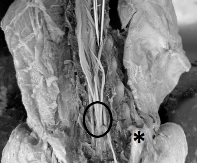
Although in one individual the spinal cord extended to L5, in all analyzed individuals, the lumbar intumescence started from the last three thoracic vertebrae, extending to the first lumbar. In the lumbosacral space, between the sixth lumbar vertebra (L6) and the sacrum, only terminal filament and extensions of the spinal nerves that went towards the regions of the pelvic limbs and tail were visualized (Fig. 2).

DISCUSSION
The spinal canal of the neotropical river otter is formed by the epidural space, the meninges, the cerebrospinal fluid, and the spinal cord (Ramsey, 1959). By definition, the epidural space is located between the dura mater and the spine. This space is also called intradural space (Groen and Ponssen, 1991). Epidural applications in small animals are commonly performed in the lumbosacral intervertebral space, as the L-S intervertebral space provides better access to the epidural environment, and closer approximation to the spinal cord (Valverde and Skelding, 2019).
Among species, there are variations in the number of lumbar vertebrae. In domestic carnivores, the number of lumbar vertebrae has an average of seven vertebrae (Sisson et al., 1975). In wild carnivores, seven vertebrae exist for example in the lion (Pantera leo) (Santos et al., 2006), in the giant otter (Pteronura brasiliensis) (Machado et al., 2009), and in South American fur seal (Arctocephalus australis) (Machado et al., 2003). These data differ from our findings of only six lumbar vertebrae in three neotropical river otters (Lontra longicaudis).
However, it is like what has been described for the tayra (Eira barbara) (Branco et al., 2013; Adami et al., 2015), the maned wolf (Chrysocyon brachyurus) (Machado et al., 2002), jaguarundi (Herpailurus yagouaroundi) (Carvalho et al., 2003) and the South American ring-tailed coati (Nasua nasua) (Gregores et al., 2010). In all these carnivore species, only six lumbar vertebrae have been described.
Also, regarding the conus medullaris, different descriptions in different animal species can be found. For example, the medullary conus in the spinal cord of the tayra ends at the level of the fifth and sixth lumbar vertebrae, in the giant otter this occurs at the level of the fourth lumbar vertebra (Machado et al., 2009; Branco et al., 2013).
The results for the giant otter were similar to what was verified during this study in the neotropical river otter. In two animals (A and B) the conus medullaris ended at the level of the fourth lumbar vertebra. However, in one animal (C) it ended at the fifth lumbar vertebra. At the level of the sixth lumbar vertebra the spinal cord segments were not observed as mentioned for the tayra (Branco et al., 2013; Adami et al., 2015). Here, only extensions of spinal nerves could be seen.
In domestic carnivores like dogs and cats, variations are also known. In dogs, the medullary conus can reach up to the level of L7, and in cats the spinal cord reaches the sacral vertebrae (Evans and Lahunta, 2012; Dyce et al., 2019). In domestic animals, a huge amount of literature exists about spinal cord morphology and anatomy. This is especially helpful regarding anesthetic procedures and surgery procedures.
Regarding anesthesia, epidural anesthesia in veterinary practice is mostly used in large animals like cows, horses and sheep. However, it is also described in dogs. For example, more than half a century ago it was described in dogs by Bone and Peck (Bone; Peck, 1956). And according to Martin-Flores (2019), the epidural space at the lumbosacral level is the target location for an epidural injection. The space between the sacrum and the first caudal (sacrococcygeal) vertebra can also be used (Massone, 2019). The results of our anatomical study show, that the same locations theoretically could be used in the neotropical river otter.
Considering the results like those found by Machado et al. (2009) for giant otters, the lumbosacral level can also be used for epidural anesthesia in this species. However, for the tayra, the authors suggested the sacrococcygeal region as the choice for the use of this anesthesia (Branco et al., 2013), although in cats, which present a medullary cone arrangement beyond the last lumbar vertebra, the lumbosacral level is also indicated for epidural anesthesia in this species (Martin-Flores, 2019; Massone, 2019).
Epidural anesthesia represents a popular technique of local anesthesia in veterinary medicine (Valverde, 2008). For a safe anesthetic procedure, it is necessary that the painful sensation be controlled and allow the performance of surgical interventions without suffering to the patient (Silva, 2003). Epidural anesthesia is considered a safe technique when compared to general anesthesia, as it reduces intraoperative stress and minimizes the risks of anesthetic intervention, being widely used in critically ill and elderly patients (Martin-Flores, 2019). According to Massone (2019), epidural anesthesia causes a blockage in the sensory and motor roots of the spinal nerves due to the deposition of local anesthetic around the Dura mater, which diffuses into the epidural space.
The decision which kind of anesthetic method is the best for a patient depends, of course, on several different points which involve, for example, the patient, the surgery procedure, and the abilities of the veterinarian. Therefore, it has to keep in mind that this is an anatomical descriptive study and not a practical trial on living animals regarding epidural anesthesia. Especially in wild animals like otters, whose movements are very fast and flexible, and restrain procedures are difficult and very stressful, epidural anesthesia might be only an additional tool and definitely not the standard anesthetic method. Anesthetic methods of choice in otters are still injectable and/or inhalation anesthesia (personal communication, Weber 2022).
CONCLUSIONS
In two animals (A and B) of neotropical river otter (Lontra longicaudis), the conus medullaris ended at the level of the fourth lumbar vertebra. However, in one animal (C) it ended at the fifth lumbar vertebra. At the level of the sixth lumbar vertebra, the spinal cord segments were not observed. Here, only extensions of spinal nerves could be seen. Similar to other animals, the space between the sacrum and the first caudal (sacrococcygeal) vertebra can theoretical be used in neotropical river otters for epidural anesthesia injection.
Acknowledgements: We want to thank Muhammad Akbar, Hahafi Ma’ruf, Team Verteb Andalas University, and all Museum Zoology Andalas University members. This study was funded by Andalas University (contract number T/49/UN.16.17/PT.01.03-IS-RD/2020).
REFERENCES
Adami, M., Rekowsky, B.S.S., Silva, R.D.G., Faria, M.M.M.D., Pinto, M.G.F., Almeida, A.E.F.S. (2015). Topografia vertebromedular de irara (Eira barbara Linnaeus, 1758). Pesq. Vet. Bras. 35(10): 871-874. http://dx.doi.org/10.1590/S0100-736X2015001000009
Barbosa, P.M.L., Esteves, P.S., Santos, A.L., Carvalho Junior, O. (2021). Vascularization of the Aortic Arch in Neotropical Otter (Lontra longicaudis, Olfers 1818). IUCN Otter Spec. Group Bull., 38: 173–182. https://www.iucnosgbull.org/Volume38/Barbosa_et_al_2021.html
Bone J.K, Peck J.G. (1956). Epidural anesthesia in dogs. J Am Vet Med Assoc. 128: 236 - 238.
Branco, E., Lins, F.L.M.L., Pereira, L.C., and Lima, A.R. (2013). Topografia do cone medular da irara (Eira barbara) e sua relevância em anestesias epidurais. Pesquisa Veterinária Brasileira, 33(6): 813-816
https://www.scielo.br/j/pvb/a/9V4LsQYz4ZyMrzbVVFfjB7C/?format=pdf
Carvalho S.F.M., Santos A.L.Q., Avila Junior R.H., Andrade M.B., Magalhães L.M., Moraes F.M., Ribeiro P.I.R. (2003). Topografia do cone medular em um gato mourisco, Herpailurus yagouaroundi (Severtzow, 1858). Archs Vet. Sci. 8: 35-38. http://dx.doi.org/10.5380/avs.v8i2.4031
Carvalho Junior, O., Barbosa, P. M. L., Birolo, A.B. (2021). Status of conservation of Lontra longicaudis (Olfers, 1818) (Carnivora: Mustelidae) on Santa Catarina Island. IUCN Otter Spec. Group Bull., 38(4): 186–201. https://www.iucnosgbull.org/Volume38/Carvalho-Junior_et_al_2021.html
Carvalho-Junior, O., Birolo, A.B., Macedo-Soares, L. (2010). Ecological Aspects of Neotropical Otter (Lontra longicaudis) in Peri Lagoon, South Brazil. IUCN Otter Spec. Group Bull., 27(2), 105–115. https://www.iucnosgbull.org/Volume27/Carvalho-Junior_et_al_2010b.html
Dyce, K.M., Sack, W.O., Wensing, C.J.G. (2019). Textbook of Veterinary Anatomy (5o edição). Elsevier Science Health Science Division.
Evans, H.E., Lahunta, A.D. (2012). Miller’s Anatomy of the Dog (4a edição). W.B. Saunders Company.
Gregores, B.G.., Branco, É, Carvalho, A.F. de, Samento, C.A.P., Oliveira, P.C., Ferreira, G., Cabral, R., Fioretto, E.T., Miglino, M. A., and Cortopassi, S.R.G. (2010). Topografia do cone medular do quati (Nasua nasua Linnaeus, 1766). Biotemas, 23 (2): 173-176 Link
Groen, R.J.M., Ponssen, H. (1991). Vascular anatomy of the spinal epidural space: considerations on the etiology of the spontaneous spinal epidural hematoma. Clin Anat 4: 413–20.
International Committee on Veterinary Gross Anatomical Nomenclature ICVGAN (2017). Nomina Anatomica Veterinaria, Sixth edition.
https://www.wava-amav.org/downloads/nav_6_2017.zip
Machado G.V., Fonseca C.C., Das Neves M.T.D., De Paula T.A.R., Benjamin L.A. (2002). Topografia do cone medular no lobo-guará (Chrysocyon brachyurus Illiger, 1815), Revta Bras. Ciênc. Vet. 9:107-109. https://periodicos.uff.br/rbcv/article/view/7548/5832
Machado G.V., Lesnau G.G., Birck A.J. (2003). Topografia do cone medular no lobo marinho (Arctocephalus australis Zimmermman, 1783). Arqs Ciênc. Vet. Zool. 6: 11-14. https://ojs.revistasunipar.com.br/index.php/veterinaria/article/view/787/687
Machado, G.V., Rosas, F.C.W., Lazzarini, S.M. (2009). Topografia do cone medular na ariranha (Pteronura brasiliensis Zimmermann, 1780). Ciência Animal Brasileira / Brazilian Animal Science, 10(1): 301–305. https://revistas.ufg.br/vet/article/view/4093
Martins-Flores, M. (2019). Epidural and Spinal Anesthesia. Vet Clin Small Anim. 49(6): 1095-1108. https://doi.org/10.1016/j.cvsm.2019.07.007
Massone, F. (2019). Anestesiologia Veterinária-Farmacologia e técnicas (7a edição). Guanabara Koogan.
Ramsey H.J. (1959). Fat in the epidural space in young and adult cats. Am J Anat. 104: 345–79. https://doi.org/10.1002/aja.1001040303
Rodrigues, L. de A., Leuchtenberger, C., Kasper, C.B., Carvalho Junior, O., de Oliveira, S., Fonseca, V. (2013). Avaliação do risco de extinção da Lontra neotropical Lontra longicaudis (Olfers, 1818) no Brasil. Biodiversidade Brasileira, 3(1): 216–227. https://revistaeletronica.icmbio.gov.br/BioBR/article/view/389/334
Santos, A.L.Q., Silva, J.M.N., Kaminishi, A.P.S, Gomes, D.D., Vieira, L.G, Hirano, L.Q.L., Pereira, P.C., Cintra, R.V., Brito, F.M.M., Bosso, A.C.S., Ferreira, C.G. (2006). Estudo anatômico das vértebras lombares do leão (Panthera leo Linnaeus, 1758) relato de caso. Vet. Not. 12(2). https://seer.ufu.br/index.php/vetnot/article/view/18739
Silva, O.C., Marques, J.A. (2003). Epidural analgesia in cows through the use of morphine and lidocaine combined. Ars Veterinaria, 19(1): 21–25.
Sisson, S., Grossman, J.D., Getty, R. (1975). Sisson and Grossman’s The Anatomy of the Domestic Animals.: Vol. 1,2 (5th Edition). W B Saunders Co. ISBN 9780721641027, 9780721641072, 0721641024, 0721641075 https://www.worldcat.org/title/Sisson-and-Grossman%27s-The-anatomy-of-the-domestic-animals/oclc/1057726
Wilson, D.E., Reeder, D.A.M. (Eds.) (2005). Mammal species of the world: a taxonomic and geographic reference, 3rd ed. Johns Hopkins University Press, Baltimore.
Valverde, A. (2008). Epidural analgesia and anesthesia in dogs and cats. Vet. Clin. N. Am. Small Anim. Pract. 38: 1205–1230. https://doi.org/10.1016/j.cvsm.2008.06.004
Valverde A, Skelding A. (2019). Comparison of calculated lumbosacral epidural volumes of injectate using a dose regimen based on body weight versus length of the vertebral column in dogs. Vet Anaesth Analg. 46: 135–40. https://doi.org/10.1016/j.vaa.2018.10.002
Weber H. (2022). personal communication.
Résumé: Étude Anatomique du Conus Medullaris chez la Loutre à Longue Queue (Lontra longicaudis)
La morphologie et la physiologie de Lontra longicaudis ne sont pas bien connues, ce qui rend parfois difficile la compréhension de son anatomie ou de son comportement. Une étude anatomique descriptive de la moelle épinière des loutres à longue queue est présentée ici. Elle a été menée au Laboratoire de Recherche de la Faune sauvage de l’Université Fédérale de Santa Catarina au Brésil. L’Institut Ekko au Brésil (IEB) – Projet Loutre a utilisé trois loutres femelles, deux loutrons et un adulte. Pour étudier la moelle épinière et ses composants, des mesures de la longueur du corps, de la musculature épiaxiale et des arcs vertébraux ont été réalisées entre la transition cervico-thoracique et la base de la queue. Le cône médullaire atteint la quatrième vertèbre lombaire. Chez un individu, il a atteint la cinquième vertèbre lombaire. Au niveau de la sixième vertèbre lombaire, aucun segment de moelle épinière n'a été observé. Chez les loutres à longue queue, l’espace entre le sacrum et la première vertèbre caudale (sacrococcygienne) peut être utilisé pour l’injection d'une anesthésie péridurale. Les praticiens doivent prendre en compte cette description lors de l’examen des zones d’injection pour une anesthésie péridurale.
Revenez au dessus
Resumen: Estudio Anatómico del Conus Medullaris en la Nutria Neotropical (Lontra longicaudis)
Las nutrias son los predadores tope en los humedales. Las nutrias tienen un rol ecológico esencial en la preservación de la La morfología y fisiología de Lontra longicaudis no son bien comprendidas, lo que en ocasiones hace difícil entender su anatomía ó comportamiento. Presentamos un estudio anatómico descriptivo de la médula espinal de la nutria neotropical. Fue conducido en el Laboratorio de Investigación en Animales Silvestres de la Universidad Federal de Santa Catarina, Brasil. Se utilizaron tres nutrias hembra (dos crías y un adulto), oriundos del Instituto Ekko Brasil (IEB) - Proyecto Nutria. Para analizar la médula espinal y sus componentes, registramos las mediciones de largo corporal y de la musculatura epiaxial y los arcos vertebrales, desde la transición cérvico-torácia hasta la base de la cola. El conus medullaris alcanzó hasta la cuarta vértebra lumbar. En un animal, alcanzó hasta la quinta vértebra lumbar. Al nivel de la sexta vértebra lumbar, no fueron observados segmentos de médula espinal. En las nutrias neotropicales, el espacio entre el sacrum y la primera vértebra caudal (sacrococcígea) puede ser usado para inyección de anestesia epidural. Los que deben intervenir animales deberían considerar ésta descripción al considerar los puntos de inyección para anestesia epidural.
Vuelva a la tapa
Resumo: Estudo Anatômico do Conus Medular em Lontra Neotropical (Lontra longicaudis)
La morfologia e fisiologia do Lontra longicaudis não são bem compreendidas, o que, às vezes, dificulta compreender a sua anatomia ou comportamento. Um estudo anatômico descritivo da medula espinhal de lontras neotropicais é apresentado. A pesquisa foi conduzida no Laboratório de Pesquisa em Animais Selvagens da Universidade Federal de Santa Catarina, Brasil. Foram usadas várias fêmeas adultas e dois filhotes de lontras, oriundos do Instituto Ekko Brasil (IEB) — Projeto Lontra. Para analisar a medula espinhal e seus componentes, foram coletadas as medidas do comprimento corporal e da musculatura epiaxial e arcos vertebrais desde a transição cervicotorácica até a base da cauda. O cone medular atingiu a quarta vértebra lombar. Em um animal, atingiu a quinta vértebra lombar. Ao nível da sexta vértebra lombar não foram observados segmentos da medula espinhal. Em lontras de rio neotropicais, o espaço entre o sacro e a primeira vértebra caudal (sacrococcígea) pode ser usado para injeção de anestesia peridural.
Voltar ao topo


