IUCN/SSC Otter Specialist Group Bulletin

©IUCN/SCC Otter Specialist Group
Volume 41 Issue 1 (March 2024)
Citation: Fujita, E.S., Mello, D.M.D. De, Nascimento Filho, A.R. Do, Fonseca-Alves, C.E., Tochetto, C., and Rosas, F.C.W. (2024). Metastatic Mammary Gland Carcinoma in a Giant Otter (Pteronura brasiliensis). IUCN Otter Spec. Group Bull., 41 (1): 53 - 62
Metastatic Mammary Gland Carcinoma in a Giant Otter (Pteronura brasiliensis)
Estefani Segato Fujita1, Daniela Magalhães Drummond De Mello2,3*, Antony Rodrigues Do Nascimento Filho2, Carlos Eduardo Fonseca-Alves4, Camila Tochetto5, and Fernando C. Weber Rosas2
1Instituto de Desenvolvimento Sustentável Mamirauá - IDSM. Escritório de Projetos, Tefé, Amazonas, Brazil
2Aquatic Mammals Laboratory, National Institute for Amazonian Research, Manaus, Amazonas, Brazil
3Institute of Biosciences, University of São Paulo, São Paulo, Brazil
4School of Veterinary Medicine and Animal Science, São Paulo State University, Botucatu, São Paulo, Brazil
5Ministério da Defesa, Exército Brasileiro. 13ª CIA DAM, Itaara, Rio Grande do Sul, Brazil.
*Corresponding Author Email: danielamello@hotmail.com
Received 4th October 2023, accepted 16th October 2023
Abstract: Diagnostics of neoplasia in otters kept in captivity have been discreetly increasing over the last years, especially in older individuals. Here, we describe a case of a mammary gland carcinoma in a nulliparous 17-year-old female giant otter (Pteronura brasiliensis) kept at the National Institute for Amazonian Research in Brazil. It represents the first report of mammary carcinoma in a giant otter. The animal presented with lameness of the right hind limb, weight loss, dyspnea, impaired locomotion, and signs of pain. Physical examination was performed with the observation of a medium sized mass of approximately 5 cm3 in the left mammary gland. On that occasion, samples for cytology and blood tests were collected. Results suggested the presence of mammary carcinoma and health alterations. The individual showed progressively poor health and was non-responsive to medical treatment. Euthanasia was performed and necropsy showed cachexia, a nodule in the mammary gland and an increase in the volume of mesenteric lymph nodes. Nodules from the mammary gland, pulmonary parenchyma, and mesenteric lymph node were collected for histopathological and immunohistochemistry analyses. Histopathology results revealed a mammary carcinoma complex subtype at stage II, with confirmed results by immunohistochemistry, which yielded positive results for CK Pan, CK19 and CK7 tumoral markers in the neoplastic cells. The regular health monitoring of captive otters may aid in the understanding of the prevalence and the etiology of this type of tumor, as also to take preventive measures to avoid premature death of individuals.
Keywords:cytology, immunohistochemistry, histopathology, neoplasia, otter
INTRODUCTION
The occurrence and diagnostics of neoplasia in aquatic mammals kept in captivity have been increasing in the past few years. The reasons behind that may be related to the increasing ages of animals maintained in captivity, hence higher probability of developing neoplasia. Besides that, the increased number of pathologists and more effective diagnostic tools allowed for more numerous and precise detection of neoplasia (Colegrove, 2018). Despite of that, the identification and diagnose of neoplasia in otters remain deficient. Different types of neoplasia have been diagnosed in only seven out of 13 otter species (Reimer and Lipscomb, 1998; Nakamura et al., 2002; Bae et al., 2007; Pye et al., 2010; Stedman and Mills, 2014; Van De Velde et al., 2019; Pinto et al., 2023). In most cases, spontaneous tumors of unknown etiology were described (Bae et al., 2007; Burek-Huntington et al., 2012; Tanaka et al., 2013); while in some cases carcinogenic chemicals have been related as potential causes of neoplasia in free-ranging otters (Rodriguez-Ramos Fernandez et al., 2012). Frequently, the tumors are associated to secondary infectious viral and parasitic diseases due to debilitation and stress (Reimer and Lipscomb, 1998; Hsu and Mathura, 2018).
As top predators, aquatic and semi-aquatic mammals are considered sentinels of the environmental health as their overall health ultimately reflects the health of the ecosystems upon which they inhabit (Burek et al., 2008; Moore, 2008). The contact with carcinogenic agents may lead to the development of a variety types of neoplasia, including those related to the reproductive tract including mammary glands (Martineau et al., 2002).
Mammary cell proliferation has been extensively described in domestic dogs and cats (Rauner et al., 2018) and less frequently described in zoo canids (Munson and Moresco, 2007) and other carnivore species such as browns bears (Vashist et al., 2013) and carnivore marsupials (Canfield et al., 1990). On the other side, mammary tumors represent a major concern in wild felines representing the most frequent reproductive tumors diagnosed and commonly metastasized widely (Kloft et al., 2019). So far, one case of metastatic mammary carcinoma has been described in otter - the Asian small-clawed otter (Aonyx cinereus) (Muller et al., 2020). A tubular adenocarcinoma with a distinct stromal component and lymph node metastasis were diagnosed in the excised mammary tissue 9 months prior to the euthanasia of the 11 years old otter (Muller et al., 2020). Mammary carcinomas, as well as other types of tumors in otters, seems to be more frequently reported in captive adult individuals.
CASE REPORT
A 17-year-old nulliparous female giant otter (P. brasiliensis), maintained at the National Institute of Amazonian Research (INPA) was observed presenting asthenia, hyporexia, hypodipsia, and dyspnea in January 2016. Physical examination was performed after immobilization with Zolazepam (1.5 mg kg-1) and Acepromazine (0.1 ml kg-1) followed by an injection of atropine sulfate 1 % (0.09 mg kg-1) (Rosas et al., 2008). Heart rate, respiratory frequency and body temperature were monitored during the anesthetic procedure in a room with controlled temperature (23 °C). All physical parameters were within normal ranges for the species (Table 1).
| Table 1: Cardiac rate, respiratory frequency, and body temperature of one female giant otter under 89 min of general anesthesia. | |||
| Pteronura brasiliensis1 | Pteronura brasiliensis2 | Lontra longicaudis2 | |
| Zolazepam + Acepromazine | Ketamine Hydrochloride | ||
| Cardiac rate (beats/min) | 88 - 129 | 115 | 179 -186 |
| Resp. frequency (breaths/min) | 28 - 68 | ND | ND |
| Rectal temperature (˚C) | 37 - 39.5 | 39.4 | 39.3 - 40.8 |
| 1 Present study 2 Marsicano et al. (1986) ND = not determined |
|||
Blood was drawn from the femoral vein for hematological and serum chemistry analyses. A medium size mass was of approximately 5 cm3 was observed in the left mammary gland during the physical examination. The mass was firm, with irregular borders, adhered to the adjacent tissue, not encapsulated, and composed by nodules of different sizes, with the largest of approximately 4-5 cm. A fine-needle aspiration cytology was performed on the mammary gland nodule to obtain a cell sample.
RESULTS
Most of the blood values showed abnormal values falling outside the normal range for the species. While the red cell series was mostly withing the normal range, the absolute and relative white blood cell counts showed values that ranged from discrete to severe alteration (Table 2).
| Table 2: Complete blood count and serum chemistry of a giant otter (P. brasiliensis) with mammary gland adenocarcinoma. | ||
| Complete Blood Count | This study | Reference Range (Rosas et al., 2008) |
| RBC (106 mm3 -1) | 5.3 | 5.3 - 8.6 |
| Hematocrit (%) | 42.0 | 41.3 - 62.6 |
| Hemoglobin (g dL-1) | 15.3 | 13.9 - 21.2 |
| MCV (fL) | 79.7 | 65.0 - 82.5 |
| MCHC (g dL-1) | 36.4 | 32.0 - 33.9 |
| Platelets (103 mm3 -1) | 390.0 | 198.0 - 619.5 |
| White Blood Count (103 mm3 -1) | 8.8 | 2.8 - 7.4 |
| Neutrophils (%) | 76.0 | 74.0 - 83.0 |
| Eosinophils (%) | 12.9 | 0.0 - 2.30 |
| Lymphocytes (%) | 2.0 | 13.7 - 22.0 |
| Monocytes (%) | 10.0 | 0.8 - 1.5 |
| Basophils (%) | 0.0 | 0.0 - 0.7 |
| Serum chemistry | ||
| ALT (UI L-1) | 40.0 | 58.0 - 209.0 |
| AST (UI L-1) | 120.0 | 142.0 - 223.0 |
| Total protein (g dL-1) | 9.0 | 6.9 - 7.1 |
| Creatinine (mg dL-1) | 5.1 | 1.1 - 2.8 |
| Alkaline Phosphatase (UI L-1) | 87.8 | 40.0 - 77.0 |
| Blood Urea Nitrogen (mg dL-1) | 267.0 | 99.0 - 344.0 |
The cytology showed predominantly epithelial cells, in groups or isolated, with cytoplasmic vacuoles, lack of pigments, and few mesenchymal cells. A large number of cells showed more than three characteristics of malignancy: Anisocytosis, anisokaryosis, multinucleate cells, irregular chromatin distribution within nuclei and evidence of mitotic figures. Results compatible with mammary carcinoma. Fine needle aspiration cytology and blood work confirmed the clinical observations.
Supportive treatment care was performed with IV fluids (Lactated Ringer’s Solution) and antibiotics (Cefovecin, 8 mg kg-1). Ten days after the initial treatment, the animal was non-responsive and even more debilitated. At this point, euthanasia was performed. Necropsy showed cachexia, a nodule in the mammary gland and increase of volume of mesenteric lymph nodes. The tissues were fixated by immersion in neutral buffered 10% formalin for histopathological and immunohistochemistry analyses.
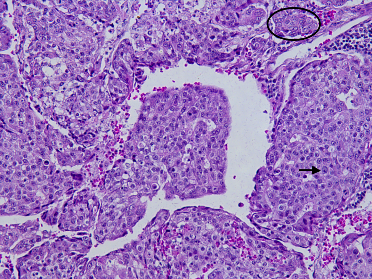
The histopathological examination revealed a solid carcinoma (Fig. 1), characterized by partially encapsulated and infiltrative neoplasm, tumor cells were arranged in solid sheets, cords, or nests. The neoplastic cells exhibited moderate pleomorphism, anisocytosis and anisokaryosis. Some binucleate cells were observed. The cytoplasmic boundaries were moderately distinct, the cytoplasm was moderate and slightly eosinophilic. Chromatin was poorly aggregated and 1-3 conspicuous nucleoli were observed. There were 2-5 mitosis figures per field of highest magnification. The stroma was characterized by fibrous connective tissue, which varies from scarce to abundant, in some places, where it is arranged in multidirectional bundles. Areas of necrosis and mineralization were also observed. There were neoplastic cells inside lymphatic and lymphatic vessels. Metastasis was observed in the mesenteric lymph nodes, whose parenchyma was replaced by neoplastic cells.
Immunohistochemistry revealed neoplastic cells negative for tumoral markers Vimentin and CK20, and positive for markers CK Pan, CK19, and CK7, supporting the metastatic mammary adenocarcinoma diagnosis.

DISCUSSION
Wild animals have been considered environmental sentinels since the Roman Era due to the sensitivity of some species to environmental changes. They provide information such as type, characteristics, quantity, and prevalence of pollutants as well as their environmental effects (Fox, 2001). Therefore, human, animal and environmental health are directly connected (Zinsstag et al., 2011). A large amount of biological material from wildlife species comes from necropsies, and the information gained from postmortem examinations animals kept in rehabilitation and exhibition facilities are crucial for a better understanding of the relation between their health and the environment. Moreover, the possibility of having a compilation of the medical history from captive individuals forms the basis of understanding a certain disease and its impact at the individual, population, species, and even ecosystem level (McAloose et al., 2018).
According to Bostock (1986), the mammary carcinoma has a complex etiology with a high level of malignancy and high metastatic capacity. Animals with mammary carcinomas can show a very rapid clinical decline, as noticed in this case. The observed leukocytosis accompanied by increased values of monocytes and eosinophils have demonstrated the neoplastic and inflammatory stimulus of the mammary neoplasia. The blood alterations shown in Table 2 keep up with the clinical signs observed, together with metabolic dysfunction, alteration of organic functions and a compromised immune system. The histopathological results were similar to what was previously described in the literature for other mammals with mammary cancer (Carpenter et al., 1980; Bryant et al., 2007; Cassali et al., 2007; Munson and Moresco, 2007).
In otters, five different types of carcinomas have been reported so far. Most of them in animals kept in captivity and over eight years old. A differentiated basal cell carcinoma was found in the left submandibular region of a captive Cape clawless otter (Aonyx capensis) of > 8 years (Nakamura et al., 2002). Cholangiocellular adenocarcinoma was described in an adult female captive sea otter (Enhydra lutris) (Stetzer et al., 1981). A primary pleural squamous cell carcinoma was found in a free-ranging North American river otter (Lontra canadensis) (Van De Velde et al., 2019), representing the only carcinoma described in a free-ranging otter species. A thyroid gland carcinoma was diagnosed in a 20 years old captive small-clawed otter (Aonyx cinereus) (Hsu and Mathura, 2018). In other occasion, one case of metastatic exocrine pancreatic adenocarcinoma was described in an 18-year-old giant otter (Pteronura brasiliensis) in captivity with a history of inappetence and apathy (Pinto et al., 2023). All the individuals above cited were adults, reinforcing the importance of close monitoring of health from otters kept in captivity, especially when dealing with endangered species such as giant otter (Groenendijk et al., 2021).
CONCLUSION
This study is the first case report on mammary carcinoma in P. brasiliensis. The regular health monitoring of confined animals combined with epizootiological and pathological studies are necessary to determine the prevalence, and the etiology of this type of tumor in giant otters, as well as to take preventive measures to avoid premature death of individuals.
Acknowledgments - We thank the Conselho Nacional de Desenvolvimento Científico e Tecnológico -CNPq, Associação dos Amigos para a Proteção do Peixe-boi da Amazônia – AMPA (Projeto Mamíferos Aquáticos da Amazônia sponsored by Petrobras Socioambiental) for logistical and/or financial support, and Rodrigo Amaral for technical support.
REFERENCES
Bae, I.-H., Pakhrin, B., Jee, H., Shin, N.-S., Kim, D.-Y. (2007). Hepatocellular adenoma in a Eurasian otter (Lutra lutra). Journal of Veterinary Science, 8(1): 103 -105. https://doi.org/10.4142/jvs.2007.8.1.103
Bostock, D. E. (1986). Veterinary professional development series canine and feline mammary neoplasms. British Veterinary Journal, 142(6): 506–505. https://doi.org/https://doi.org/10.1016/0007-1935(86)90107-7
Bryant, B., Portas, T., Montali, R. (2007). Mammary and pulmonary carcinoma in a dromedary camel (Camelus dromedarius). Australian Veterinary Journal, 85(1–2): 59–61. https://doi.org/10.1111/j.1751-0813.2006.00093.x
Burek, K. A., Gulland, F. M. D., O’Hara, T. M. (2008). Effects of climate change on arctic marine mammal health. Ecological Applications, 18(sp2): S126–S134. https://doi.org/10.1890/06-0553.1
Burek-Huntington, K. A., Mulcahy, D. M., Doroff, A. M., Johnson, T. O. (2012). Sarcomas in three free-ranging northern sea otters (Enhydra lutris kenyoni) in Alaska. Journal of Wildlife Diseases, 48(2): 483–487. https://doi.org/10.7589/0090-3558-48.2.483
Canfield, P. J., Hartley, W. J., Reddacliff, G. L. (1990). Spontaneous proliferations in Australian marsupials - a survey and review. 1. Macropods, koalas, wombats, possums and gliders. Journal of Comparative Pathology, 103: 135-146.
Carpenter, J. W., Davidson, P., Novilla, M. N., Huang, J. C. (1980). Metastatic, papillary cystadenocarcinoma of the mammary gland in a black-footed ferret. Journal of Wildlife Diseases,16(4): 587-592. https://doi.org/10.7589/0090-3558-16.4.587
Cassali, G. D., Gobbi, H., Malm, C., Schmitt, F. C. (2007). Evaluation of accuracy of fine needle aspiration cytology for diagnosis of canine mammary tumours: Comparative features with human tumours. Cytopathology, 18(3): 191–196. https://doi.org/10.1111/j.1365-2303.2007.00412.x
Colegrove, K.M. (2018). Noninfectious diseases. In: Gulland, F.M.D., Dierauf, L.A. Whitman, K.L. (Eds). CRC Handbook of Marine Mammal Medicine, 3rd Edition. CRC Press, Boca Raton, pp. 255-284. ISBN: 978-1498796873
Fox, G. (2001). Wildlife as sentinels of human health effects in the great lakes–St. Lawrence basin. Environmental Health Perspectives, 109(6): 853–861. https://doi.org/10.1289/ehp.01109s6853
Groenendijk, J., Marmontel, M., Van Damme, P., Schenck, C., Wallace, R. (2021). Pteronura brasiliensis. The IUCN Red List of Threatened Species, 2015, e.T18711A21938411. https://doi.org/10.2305/IUCN.UK.2021-3.RLTS.T18711A164580466.en
Hsu, C. Da, Mathura, Y. (2018). Severe visceral pentastomiasis in an oriental small-clawed otter with functional thyroid carcinoma. Journal of Veterinary Medical Science, 80(2): 320–322. https://doi.org/10.1292/jvms.17-0383
Kloft, H. M., Ramsay, E. C., Sula, M. M. (2019). Neoplasia in captive Panthera species. Journal of Comparative Pathology, 166: 35–44. https://doi.org/10.1016/j.jcpa.2018.10.178
Marsicano, G., Marmontel-Rosas, M., Rosas, F. C. W., Souza, R. H. S. (1986). Contençao de lontras amazônicas com Cloridrato de Ketamina [Contention of Amazonian otters with Ketamine hydrochloride]. A Hora Veterinária, 31: 5-8.
Martineau, D., Lemberger, K., Dallaire, A., Labelle, P., Lipscomb, T. P., Michel, P., Mikaelian, I. (2002). Cancer in wildlife, a case study: beluga from the St. Lawrence estuary, Quebec, Canada. Environmental Health Perspectives, 110(3): 285-292. https://doi.org/10.1289/ehp.02110285
McAloose, D., Colegrove, K. M., Newton, A. L. (2018). Wildlife necropsy. Pp. 1–20 in Terio, K.A., McAloose, D., St. Leger, J. (Eds) Pathology of Wildlife and Zoo Animals. Elsevier. ISBN: 978-0-12-805306-5 https://doi.org/10.1016/B978-0-12-805306-5.00001-8
Moore, S. E. (2008). Marine mammals as ecosystem sentinels. Journal of Mammalogy, 89(3): 534–540. https://doi.org/10.1644/07-MAMM-S-312R1.1
Muller, J., Fischer, D., von Bomhard, W., Henrich, M., Herden, C. (2020). Metastatic mammary carcinoma in an Asian small clawed otter (Aonyx cinereus). Journal of Comparative Pathology, 174: 172. https://doi.org/10.1016/j.jcpa.2019.10.108
Munson, L., Moresco, A. (2007). Comparative pathology of mammary gland cancers in domestic and wild animals. Breast Disease, 28: 7–21. https://doi.org/10.1016/j.jcpa.2019.10.108
Nakamura, K., Tanimura, H., Katsuragi, K., Shibahara, T., Kadota, K. (2002). Differentiated basal cell carcinoma in a Cape clawless otter (Aonyx capensis). Journal of Comparative Pathology, 127(2–3): 223–227. https://doi.org/10.1053/jcpa.2002.0582
Pinto, M. H. B., Braga, T. R. C., Blume, G. R., Oliveira, L. B. de, Chagas, N. T. C. das, Souza, F. R., Cassali, G. D., Sant’Ana, F. J. F. de. (2023). Metastatic exocrine pancreatic adenocarcinoma in a giant otter (Pteronura brasiliensis). Acta Veterinaria Hungarica., 71(1): 41-45 https://doi.org/10.1556/004.2023.00888
Pye, G. W., White, A., Robbins, P. K., Burns, R. E., Rideout, B. A. (2010). Preventive medicine success: thymoma removal in an African spot-necked otter (Lutra maculicollis). Journal of Zoo and Wildlife Medicine, 41(4): 732–734. https://doi.org/10.1638/2010-0068.1
Rauner, G., Ledet, M. M., Van de Walle, G. R. (2018). Conserved and variable: understanding mammary stem cells across species. Cytometry Part A, 93(1): 125–136. https://doi.org/10.1002/cyto.a.23190
Reimer, D. C., Lipscomb, T. P. (1998). Malignant seminoma with metastasis and herpesvirus infection in a free-living sea otter (Enhydra lutris). Journal of Zoo and Wildlife Medicine, 29(1): 35-39. https://www.jstor.org/stable/20095713
Rodriguez-Ramos Fernandez, J., Thomas, N. J., Dubielzig, R. R., Drees, R. (2012). Osteosarcoma of the maxilla with concurrent osteoma in a southern sea otter (Enhydra lutris nereis). Journal of Comparative Pathology, 147(2–3): 391–396. https://doi.org/10.1016/j.jcpa.2012.01.017
Rosas, F. W., d’Affonseca Neto, J. A., de Mattos, G. E. (2008). Anesthesiology, hematology and serum chemistry of the Giant Otter, Pteronura brasiliensis (carnivora, mustelidae). Arquivos de Ciências Veterinárias e Zoologia da UNIPAR, 11: 81–85. https://ojs.revistasunipar.com.br/index.php/veterinaria/article/view/2562
Stedman, N. L., Mills, Z. V. (2014). Splenic marginal zone lymphoma in an Asian small-clawed otter (Aonyx cinerea). Journal of Zoo and Wildlife Medicine, 45(3): 719–722. https://doi.org/10.1638/2014-0014R.1
Stetzer, E., T.D. Williams, and J.W. Nightingale. (1981). Cholangiocellular adenocarcinoma, leiomyoma, and pheochromocytoma in a sea otter. Journal of the American Veterinary Medical Association, 179(11): 1283-1284.
Tanaka, N., Izawa, T., Kashiwagi-Yamamoto, E., Kuwamura, M., Ozaki, M., Nakao, T., Yamate, J. (2013). Primary cerebral T-cell lymphoma in a sea otter (Enhydra lutris). Journal of Veterinary Medical Science, 75(12): 1667–1669. https://doi.org/10.1292/jvms.13-0331
Van De Velde, N., Demetrick, D. J., Duignan, P. J. (2019). Primary pleural squamous cell carcinoma in a free-ranging river otter (Lontra canadensis). Journal of Wildlife Diseases, 55(3): 728–732. https://doi.org/10.7589/2018-07-181
Vashist, V. S., Rattan, S. K., Gupta, B. B. (2013). Papillary cystadenocarcinoma of the mammary gland with metastases to the gastrointestinal tract in a Himalayan brown bear (Ursus arctos). Journal of Zoo and Wildlife Medicine, 44(2): 453–456. https://doi.org/10.1638/2011-0093R1.1
Zinsstag, J., Schelling, E., Waltner-Toews, D., Tanner, M. (2011). From “one medicine” to “one health” and systemic approaches to health and well-being. Preventive Veterinary Medicine, 101(3–4): 148–156. https://doi.org/10.1016/j.prevetmed.2010.07.003
Résumé: Courte Communication sur le Carcinome Métastatique de la Glande Mammaire chez une Loutre Géante (Pteronura brasiliensis)
Les diagnostics de néoplasie chez les loutres gardées en captivité ont augmenté discrètement ces dernières années, notamment chez les individus plus âgés. Nous décrivons ici un cas de carcinome de la glande mammaire chez une loutre géante femelle nullipare de 17 ans (Pteronura brasiliensis) gardée à l’Institut national de recherche amazonienne au Brésil. Il s’agit du premier signalement d’un carcinome mammaire chez une loutre géante. L’animal présentait une boiterie du membre postérieur droit, une perte de poids, une dyspnée, des troubles de la locomotion et des signes de douleur. L’examen physique réalisé a permis d’observer une masse de taille moyenne d’environ 5 cm3 au niveau de la glande mammaire gauche. A cette occasion, des échantillons destinés à la cytologie et des analyses de sang ont été prélevés. Les résultats suggèrent la présence d’un carcinome mammaire et une altération de l’état de santé. L’état de santé de l’individu s’est progressivement détérioré et ne répondait plus aux traitements médicaux. L’euthanasie a été réalisée et l’autopsie a montré une cachexie, un nodule dans la glande mammaire et une augmentation du volume des ganglions lymphatiques mésentériques. Des nodules de la glande mammaire, du parenchyme pulmonaire et des ganglions lymphatiques mésentériques ont été prélevés pour des analyses histopathologiques et immunohistochimiques. Les résultats histopathologiques ont révélé un sous-type complexe de carcinome mammaire au stade II, avec des résultats positifs confirmés par immunohistochimie pour les marqueurs tumoraux CK Pan, CK19 et CK7 dans les cellules néoplasiques. La surveillance régulière de la santé des loutres en captivité peut aider à comprendre la prévalence et l’étiologie de ce type de tumeur, ainsi qu’à prendre des mesures préventives pour éviter la mort prématurée des individus.
Revenez au dessus
Resumen: Carcinoma Metastásico de Glándula Mamaria en una Nutria Gigante (Pteronura brasiliensis)
El diagnóstico de neoplasias en nutrias mantenidas en cautiverio ha venido aumentando discretamente en los últimos años, especialmente en individuos más viejos. Aquí, describimos un caso de un carcinoma de glándula mamaria en una hembra nulípara de 17 años, de nutria gigante (Pteronura brasiliensis) en cautiverio en el Instituto Nacional de Investigaciones Amazónicas, en Brasil. Representa el primer reporte de carcinoma mamario en una nutria gigante. El animal presentaba cojera en el miembro trasero derecho, pérdida de peso, disnea, locomoción dificultada, y signos de dolor. Se realizó un examen físico, observando una masa de tamaño mediano, de aproximadamente 5 cm3 en la glándula mamaria izquierda. En esa ocasión, colectamos muestras para citología y pruebas sanguíneas. Los resultados sugirieron la presencia de carcinoma mamario y alteraciones en la salud. El individuo exhibió un estado de salud de deterioro progresivo, y no respondió al tratamiento médico. Se llevó a cabo eutanasia, y la necropsia mostró caquexia, un nódulo en la glándula mamaria, y un incremento en el volumen de los nodos linfáticos mesentéricos. Colectamos nódulos de la glándula mamaria, parénquima pulmonar, y nódulos linfáticos mesentéricos, para análisis histopatológicos e inmunoquímicos. Los resultados de histopatología revelaron un complejo de carcinoma mamario en estadio II, con resultados confirmados por inmunoquímica, que dieron positivos para los marcadores tumorales CK Pan, CK19 y CK7 en las células neoplásicas. El monitoreo regular de salud de las nutrias en cautiverio puede ayudar a entender la prevalencia y la etiología de éste tipo de tumor, así como a tomar medidas preventivas para evitar la muerte prematura de individuos.
Vuelva a la tapa
Resumo: Carcinoma Mamário Mestastático em Ariranha (Pteronura brasiliensis)
O diagnóstico de neoplasias em lontras mantidas em cativeiro tem aumentado ao longo dos últimos anos, especialmente em indivíduos mais velhos. No presente relato, um caso de carcinoma mamário foi descrito pela primeira vez em ariranha (Pteronura brasiliensis). Tratou-se de uma fêmea nulípara, de 17 anos de idade, mantida no Instituto Nacional de Pesquisas da Amazônia, Brasil. O animal apresentou claudicação do membro posterior direito, perda de peso, dispneia, dificuldade de locomoção e apresentava sinais de dor. Durante o exame físico, observou-se uma massa de tamanho aproximado de 5 cm³ na glândula mamária esquerda. Na ocasião, foi coletada amostra por punção aspirativa por agulha fina e sangue para citologia e testes hematológicos, respectivamente. Os resultados sugeriram a presença de um carcinoma mamário e alterações dos parâmetros sanguíneos. O animal apresentou deterioração do estado de saúde de maneira progressiva e não respondeu ao tratamento medicamentoso. Desta maneira, o indivíduo foi submetido à eunatásia e, posteriormente, à necropsia. Os achados macroscópicos demonstraram caquexia, um nódulo na glândula mamária esquerda e aumento de volume dos linfonodos mesentéricos. Amostras de tecido dos nódulos da glândula mamária, do parênquima pulmonar e dos linfonodos mesentéricos foram coletados para exame histopatológico e imuno-histoquímico. O exame histopatológico revelou carcinoma complexo grau II. Esse resultado foi confirmado por imuno-histoquímica, que demonstrou resultado positivo para os marcadores tumorais pancitoqueratina (CK Pan), CK19 e CK7 nas células neoplásicas. O monitoramento contínuo do estado de saúde de lontras mantidas em cativeiro pode servir como uma importante ferramenta para entender a prevalência e etiologia desse tipo de tumor, bem como auxiliar na tomada de medidas preventivas para evitar a morte prematura dos indivíduos.
Voltar ao início


